The segmentation of blood-vessels is an important preprocessing step for the quantitative analysis of brain vasculature. We approach the segmentation task for two-photon brain angiograms using a fully convolutional 3D deep neural network.
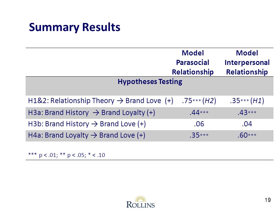
For instance, 1D centerlines can first be obtained from minimal paths and serve to initialize active contours for an accurate segmentation of the vessels’ surface (see Fig. 11). 5. Discussion. The medical interest in 3D vascular segmentation and the challenges it raises have motivated a tremendous amount of research work.This multi-step segmentation procedure consists of four steps: brain tissue segmentation (1), fuzzy-based vessel enhancement (2), extraction of cerebrovascular structures using a level-set method with anisotropic energy weights (3), and detection and correction of gaps in the vessel segmentation (4).The Vascular Modeling Toolkit. Supported by Orobix srl. vmtk is a collection of libraries and tools for 3D reconstruction, geometric analysis, mesh generation and surface data analysis for image-based modeling of blood vessels.
A review: Deep learning for medical image segmentation using multi-modality fusion. 22 Apr 2020. Due to their self-learning and generalization ability over large amounts of data, deep learning recently has also gained great interest in multi-modal medical image segmentation.
Interactive Segmentation Based on Component-trees.. Interactive 3D brain vessel segmentation from an example.. Join ResearchGate to find the people and research you need to help your work.
As each presented free example essay would, hopefully, demonstrate, good writing is a perky boat piercing briny ocean of the subject. As such, the vessel’s tonnage (read meaning-carrying capacity) should be commensurate with the demands of the waters it traverses. It also helps if your boat can float.
You can make it your own segmentation code also. Subtract the contrast filled volume with without contrast volume you will be left with the vessel and then you can easily segment out the vessels.
Research Projects. 1 A Framework for the Interactive Retrieval of Multimedia Objects. In this project we are developing an efficient and effective framework to interactively perform image segmentation and find “regions of interest” in a user input multimedia object.
This paper addresses retinal vessel segmentation on optical coherence tomography angiography (OCT-A) images of the human retina. Our approach is motivated by the need for high precision image-guided delivery of regenerative therapies in vitreo-retinal surgery. OCT-A visualizes macular vasculature, the main landmark of the surgically targeted area, at a level of detail and spatial extent.
Collaborator: Mariappan Nadar and Sandra Sudarsky, Siemens Corporate Research. Example of brain connectivity visualization. (a) A conventional method using straight lines between connected brain region. In this example, It is very difficult to see the connectivity since tons of connections.
This macro segments blood vessels in a 3D stack. It is suited for well contrasted images (low background) and works better if the width of the vessels of interest is reasonably uniform. The stack should be calibrated (micro-meter). The macro requires the ImageJ 3D suite that can be found here and this LUT (it should be copied to Fiji LUT folder).
Tubular Structure Segmentation Based on Minimal Path Method and Anisotropic Enhancement. for this work is vessel tracking in 2D and 3D images. The basic tools are minimal paths solved using the fast march-ing algorithm. This allows interactive tools for the physician.
Our Solution. True 3D renders patient-specific anatomy in an intuitive, interactive mixed reality environment. EchoPixel’s True 3D software provides an environment where clinicians can view and interact with patient specific anatomical Form, Function and Flow for improved diagnosis and more precise surgical planning.
BioMedical Engineering OnLine is an open access, peer-reviewed journal that is dedicated to publishing research in all areas of biomedical engineering. BioMedical Engineering OnLine is aimed at readers and authors throughout the world with an interest in using tools of the physical and data sciences, and techniques in engineering, to understand and solve problems in the biological and medical.
For questions related to 3D Slicer training materials and to the organization of 3D Slicer training workshops, please send an e-mail to Sonia Pujol, Ph.D., Director of Training and Education of 3D Slicer.
We demonstrate such an interactive segmentation process on the example of adaptive region growing that estimates the homogeneity criterion for segmentation and volume rendering automatically, starting from a user-specified seed point. Thereby the two subjects may be compared in the rendition. 3.1.
However, which method to select depends on the segmentation goal at hand. Not all measures are relevant for all segmentation goals. For example, if segmentation is used for brain volumetry, the overlap and volume (AVD) measures of the brain and intracranial volume (used for normalization ) segmentations are important to take into account.